

4.3 Investigating the Main Phases Nd₂Fe₁₄B and Nd₂(Fe,Co)₁₄B in NdFeB Permanent-Magnet Alloys
NdFeB permanent - magnet alloys mainly consist of three phases, namely the tetragonal phase \(Nd_{2}Fe_{14}B\), Nd - rich and B - rich phase. The \(Nd_{2}Fe_{14}B\) phase belongs to the space group P42/mm, which has been studied by Herbst, et al. The structure and properties of the B - rich phase have been studied by Givord, et al. The coercivity of NdFeB alloys has its origin in the anisotropy of \(Nd_{2}Fe_{14}B\). Therefore, the studies of the \(Nd_{2}Fe_{14}B\) phase have attracted much attention. The authors have studied the effect of Co substitution on Curie temperature by \(Nd_{2}(Fe, Co)_{14}B\) using TEM with super - high voltage of 1,000kV and Mössbauer spectroscopy (Pan, 1987; Filder, Huo, 1985; Pan, 1986; Ping, Li, Ma, Pan, et al, 1986; Ma, Jiang, Xu, 1999; Sagawa, Fujimura, fogawa, et al, 1984).
Sample Preparation and Experimental Methods for Studying Nd₂Fe₁₄B and Nd₂(Fe,Co)₁₄B
Samples of \(Nd_{15}Fe_{85 - x}B_{x}\) with \(x = 0\), 3, 5, 7 and 11 were prepared by arc melting under an Ar atmosphere. The starting materials were 99.5% Nd, 99.5% B, 99.9% Fe in purity and FeB alloy with B of 14.52 %. The ingots were remelted three times in order to achieve homogeneity. Then, the ingots were crashed and ball - milled to powders with about 3 - 5 μm. The powders were shaped in a magnetic field with 1.5 T. Finally, the samples were sintered at 1090°C and annealed at 600°C for 1 h. The prepared sample was cut into thin plate along the \(c\) axis, which was thinned to 0.025 mm by the mechanical method and followed by the ion thinned method to make the thin film for TEM experiment. TEM with super - high voltage was performed using JEM - 1000 kV. The powder samples were used in Mössbauer spectrum experiment.
SEM Analysis of NdFeB Permanent-Magnet Alloys: Microstructural Insights
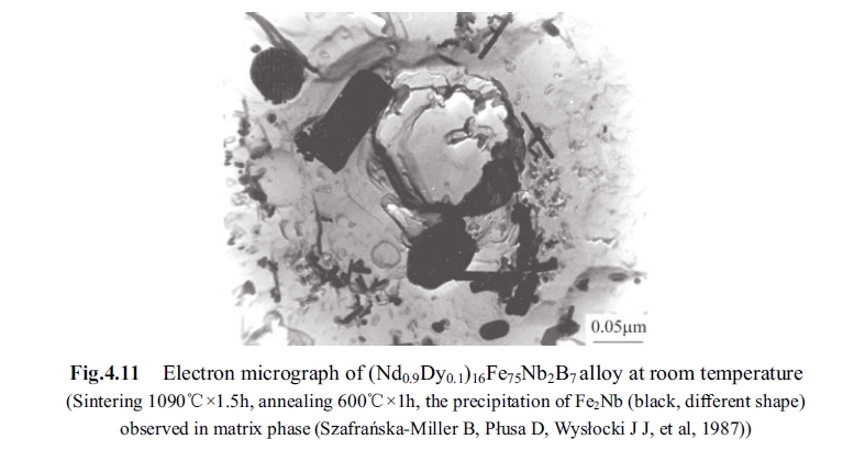
Fig. 4.11 is the SEM image of \((Nd_{0.9}Dy_{0.1})_{16}Fe_{73}Nb_{2}B_{9}\). The dark range is the tetragonal phase \(Nd_{2}Fe_{14}B\) and the white range is the Nd - rich phase. SEM with EDAX showed that the relative concentrations of Fe and Nd(Dy) are 66.62%(wt.) and 33.38%(wt.), respectively.
Formation Mechanism of the Nd₂Fe₁₄B Phase in NdFeB Magnets
In order to study the formation of \(Nd_{2}Fe_{14}B\), the samples \(Nd_{15}Fe_{85 - x}B_{x}\) with \(x = 0\), 3, 5, 7 and 11 have been prepared. X - ray diffraction showed that there are only \(\alpha - Fe\) and \(Nd_{2}Fe_{17}\) phases without B, and the new diffraction lines of \(Nd_{2}Fe_{14}B\) appear at B concentration of 3% (at.). As B>5% (at.), \(\alpha - Fe\) and \(Nd_{2}Fe_{17}\) phases completely disappear and there are main phase \(Nd_{2}Fe_{14}B\), the Nd - rich and B - rich phases. SEM with EDAX showed that the mass fractions of Fe and Nd are 65.62 % and 34.38 %, respectively, which are consistent with the fractions of Fe and Nd in \(Nd_{2}Fe_{14}B\). In conclusion, the B concentration is the decisive factor in the formation of the tetragonal phase \(Nd_{2}Fe_{14}B\). The ferromagnetic coupling of Fe and Nd is along the \(c\) axis in the tetragonal structure. The strong uniaxial anisotropy is the main cause of high coercivity for NdFeB alloys. The anisotropy has its origin in the energy splitting of rare earth ions due to the crystal field.
Room-Temperature Mössbauer Spectroscopy of Nd₂Fe₁₄B: Magnetic and Structural Analysis
The Mössbauer spectra of \(Nd_{15}Fe_{85 - x}B_{x}\) with \(x = 0\), 3, 5, 7 and 11 indicated that: at \(x = 0\), there are only the Mössbauer components of \(\alpha - Fe\) and \(Nd_{2}Fe_{17}\) phases. At \(x = 3\), the \(Nd_{2}Fe_{14}B\) appears, and at the same time, \(\alpha - Fe\) and \(Nd_{2}Fe_{17}\) still exist, as pointed by their 1st and 6th characteristic lines \(a\) and \(a'\) and by \(b\) and \(b'\), respectively. At \(x = 5\), the spectral lines of \(Nd_{2}Fe_{17}\) almost vanish and at \(x = 7\), the lines of \(\alpha - Fe\) completely disappear. There exist the sextet of the main \(Nd_{2}Fe_{14}B\), and the paramagnetic doublet of Nd - rich phase.
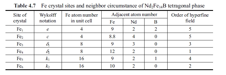
Table 4.7 lists alignment sequence of hyperfine field, which can determine sites adscription of ferromagnetic sub - spectra.
Composition Analysis and Mössbauer Spectroscopy Studies of Nd₂(Fe, Co)₁₄B
The composition analysis of electron probe has been performed for \(Nd_{2}(Fe_{1 - x}, Co_{x})_{14}B\). At \(x = 0\), the compositions of Nd and Fe are 19.4% (at.) and 84.6% (at.), respectively. The Curie temperature is 312°C; at \(x = 0.3\), the compositions of Nd, Fe and Co are 15.4% (at.), 62.5% (at.) and 22.1% (at.), respectively. Then the Curie temperature is heightened to 550°C.
In-Situ and Dynamic TEM Observations of Nd₂Fe₁₄B and Nd₂(Fe, Co)₁₄B Phases
The in situ and dynamic observation was conducted on filmy sample under electron microscope from room temperature to 130°C. At this temperature range there is no precipitate in \(Nd_{2}Fe_{14}B\), as shown in Fig. 4.12. At 260°C precipitates appeared
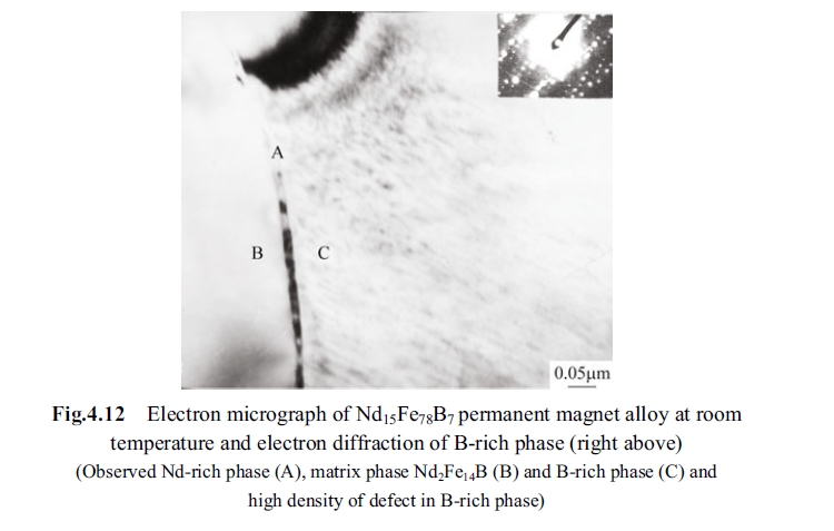
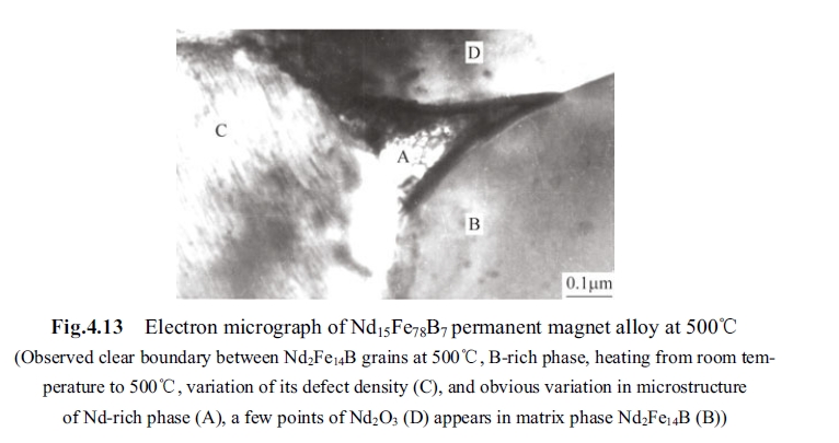
in the \(Nd_{2}Fe_{14}B\) and at 312°C highly dispersed precipitate phase was observed clearly in the all vision and diffraction ring of polycrystal appeared in diffraction pattern of \(Nd_{2}Fe_{14}B\). At 500°C more diffraction ring of polycrystal appeared, which indicated that polycrystal turned up in \(Nd_{2}Fe_{14}B\) but the crystal boundary is very clear, as shown in Fig. 4.13.
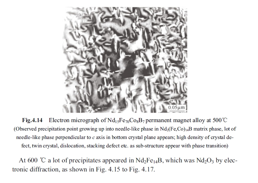
At 400°C there are tiny precipitates started to come forth in \(Nd_{2}Fe_{14}B\) and at 500°C the precipitates grew up like needles and were perpendicular to each other, such as shown in Fig.4.14.

Conclusions: Key Findings on Nd₂Fe₁₄B and Nd₂(Fe, Co)₁₄B in NdFeB Magnets
- Tetragonal crystal of \(Nd_{2}Fe_{14}B\) compound is the only hard magnetic phase. Measurement of magnetism showed \(B_{r}=1.61\ T\); by observation of TEM, the crystalline structure is intact with \(a = 0.88\ nm\), \(c = 1.22\ nm\). To enhance magnetic performance it is necessary to increase the volume fraction of the matrix phase \(Nd_{2}Fe_{14}B\) as much as possible.
- Increase of Boron content results in an increase of phase \(Nd_{2}Fe_{14}B\). When Boron content \(x\) is increased to 5, the Mössbauer spectra for \(Nd_{2}Fe_{17}\) phase is almost disappeared, and paramagnetic spectrum corresponding to B - rich phase appears synchronously.
- Increase of cobalt content leads to an enhancement of Curie temperature. The area of sub - spectra in the Mössbauer spectra reflects the comparative amount of distribution of iron atoms at crystal sites, which indirectly expresses that cobalt atom has different preferential distribution at crystal sub - sites. Analysis indicates that cobalt atoms have priority to occupy sites \(j_{2}\) and \(k_{2}\).
- Dynamic observation on filmy specimen under 1000 kV HVEM discovered that precipitate starts to appear in \(Nd_{2}Fe_{14}B\) phase at 260°C, the highly dispersed precipitate phase can be found clearly in all vision at 312°C and polycrystalline diffraction ring appears in electronic diffractive ground pattern. At 500°C more polycrystalline diffraction ring appears in diffractive ground pattern of \(Nd_{2}Fe_{14}B\).
- For \(Nd_{2}(Fe, Co)_{14}B\) phase the precipitate phase appears at 400°C, the precipitation temperature is about 100°C higher than that of \(Nd_{2}Fe_{14}B\) compound.


